Replication Stress Response
Background
We study the molecular mechanisms of the replication stress response in mammalian cells. Our overarching goal is to understand the mechanisms that ensure the stability of the genome and their involvement in cancer and ageing.
DNA replication stress is a major cause of genome instability; it is induced by DNA damaging agents such as UV, oxidants or chemotherapeutics, but can also occur spontaneously at some chromosomal regions known as replication fragile sites. A particularly relevant example of such fragile sites are telomeres: the terminal parts of our chromosomes. They have been extensively studied for their end-protection function and for the establishment of the proliferation limit, but it’s now becoming clear that these functions are strongly influenced by telomere replication. Moreover, because of their properties (location, sequence, binding proteins etc.) telomeres provide a unique context for the study of the replication stress response.
Research
We use mouse genetics, combined with newly-developed imaging techniques, including super-resolution fluorescence microscopy and electron microscopy, to analyze telomere replication at the molecular level. The main areas of study are: the role of shelterin components in telomere replication; connections between replication and telomere maintenance; the effects of exogenous stress on replication (e.g. oxidative stress or chemotherapeutics) and the molecular pathways that contribute to terminal fork stability in mammalian cells.
We are also focused in the development of new techniques and approaches that can provide the scientific community and us with new tools to tackle our biological questions.
2016.06.20
Photogallery
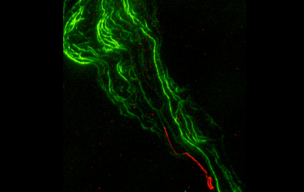 Telomeric DNA ending in a t-loop structure. Chromatin spreads, green: bulk DNA, red: telomeric DNA
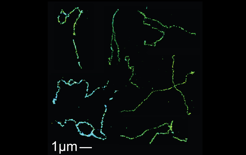 Super-resolution fluorescence microscopy imaging of telomeric DNA structures. 3D-STORM
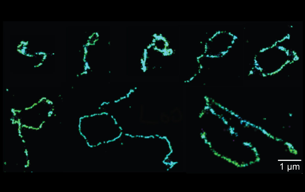 Super-resolution fluorescence microscopy imaging of telomeric t-loop structures. 3D-STORM
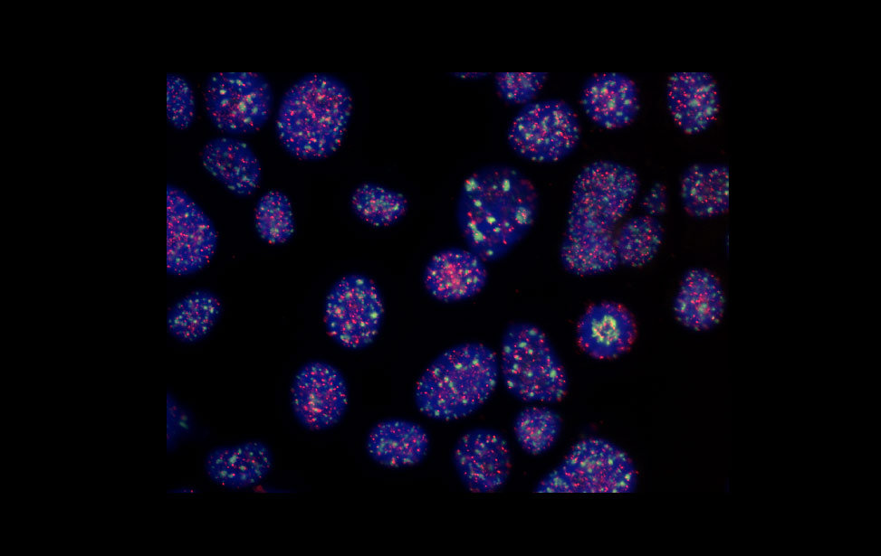 FISH-labeled mouse nuclei. Green: major satellite DNA, red: telomeres, blue: bulk DNA (DAPI)
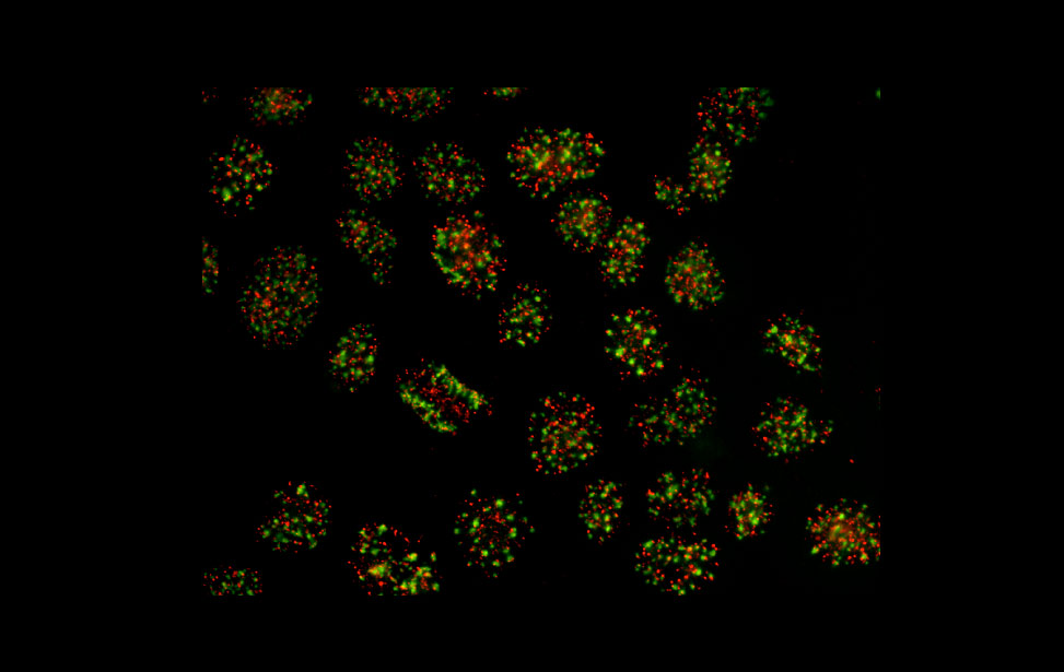 FISH-labeled mouse nuclei. Green: major satellite DNA, red: telomeres
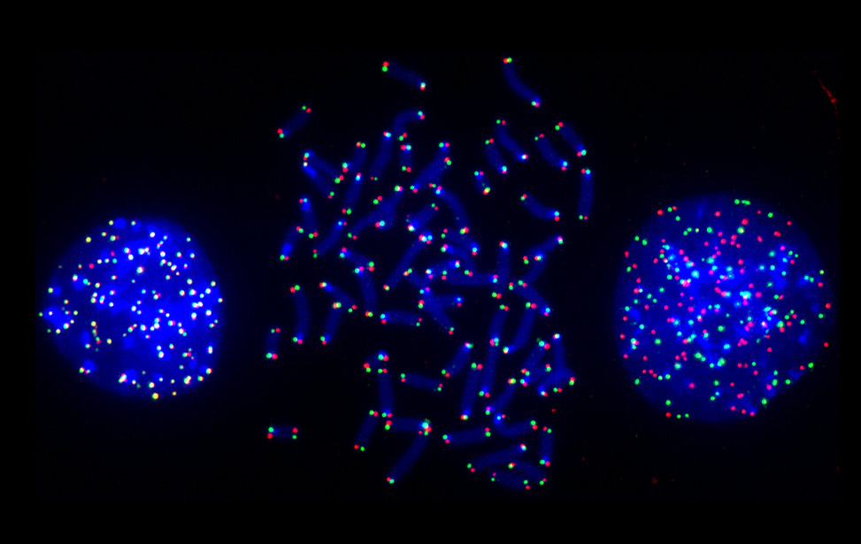 COFISH-labeling of telomeres, mouse chromosomes. Telomeres replicated by lagging strand synthesis are labeled in green while those replicated by the leading strand in red. Bulk DNA is labeled in blue.