Advanced Light Microscopy

Figure 1: Laser light tilted by a Total Internal Reflection Fluorescence Microscope (TIRFM) travelling through a fluorescence slide
Unit info
Optical imaging is a continuosly evolving field which brings more and more sophisticated visualization technologies and methods to the biomedical community, making it an essential and versatile tool for both researchers and clinicians.
Cancer research has particularly benefited from the physical properties of the light: as a matter of fact, the different ways light interacts with cells and tissues paved the way to multiple contrasts methods which are now routinely in use to explore and unravel the underlying molecular mechanisms of tumor formation and development.
The interpretation of these photophysical events and the acquisition of morphological and functional information of the relevant biological systems is possible only through synergies between different disciplines such as biology, physics, chemistry and bioinformatics.
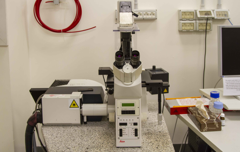
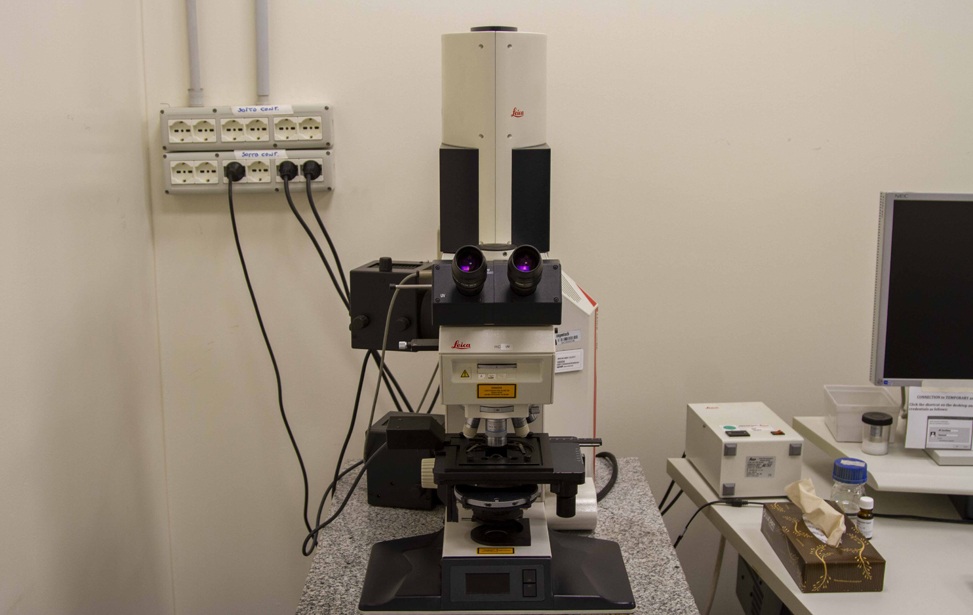
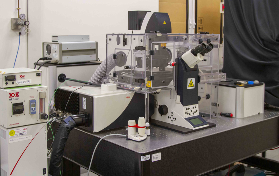
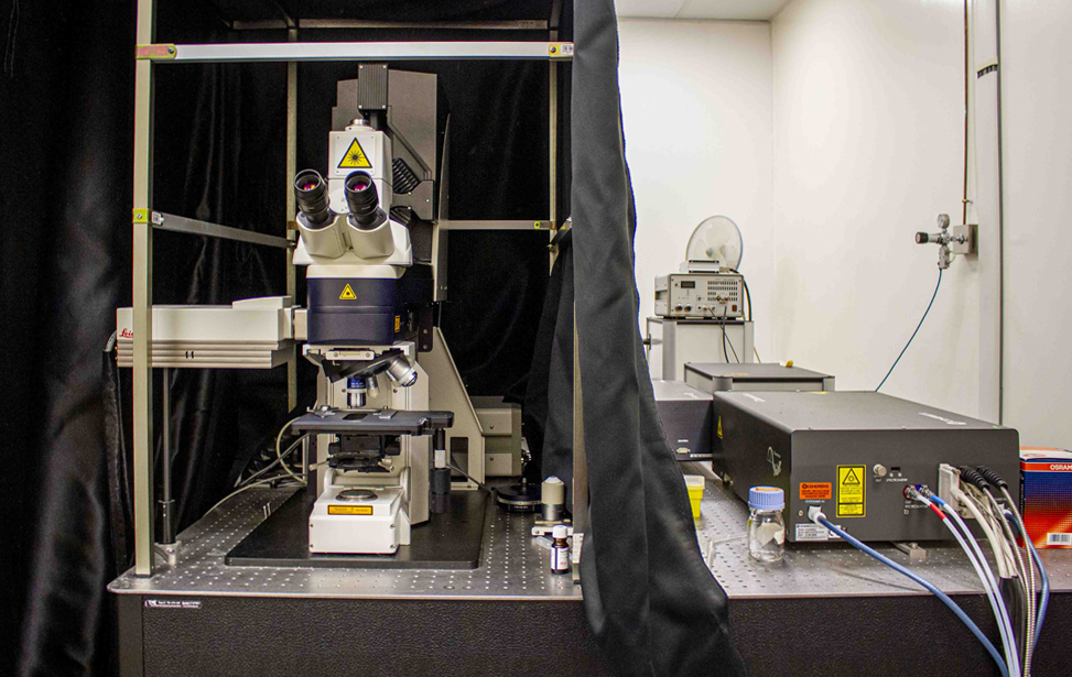
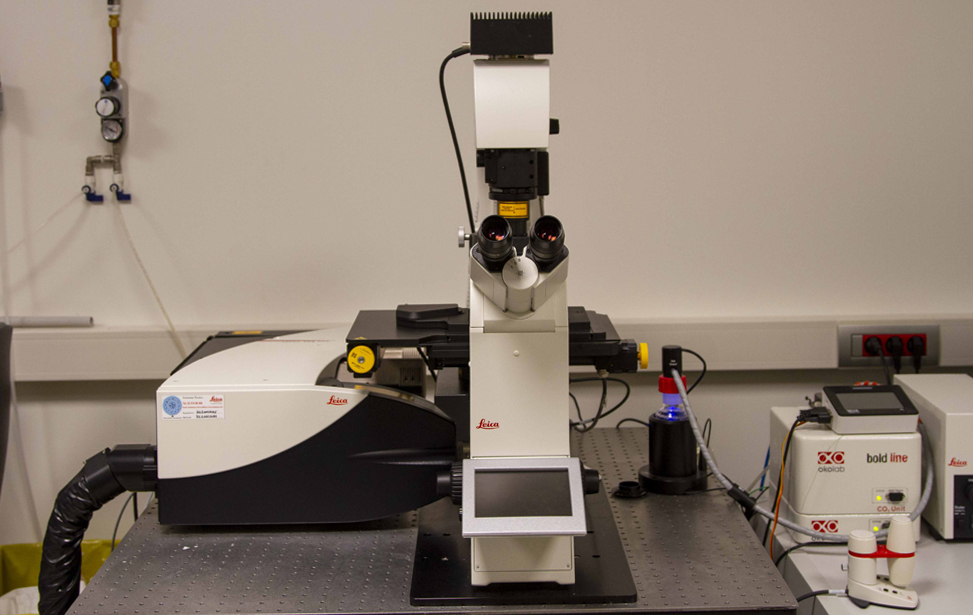
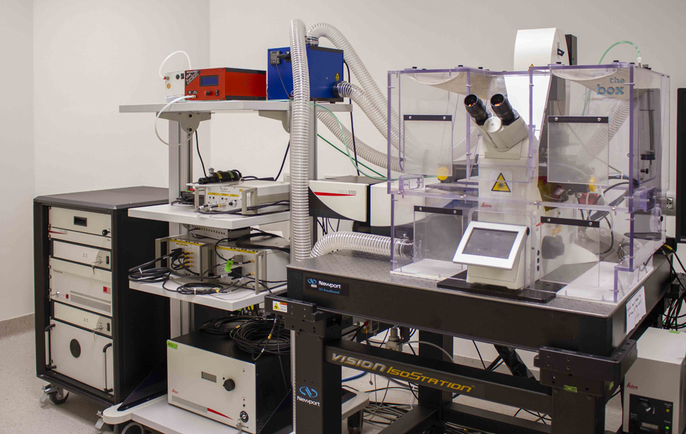
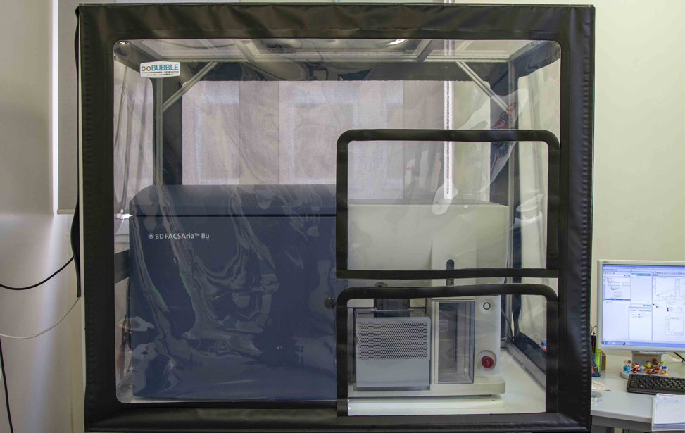
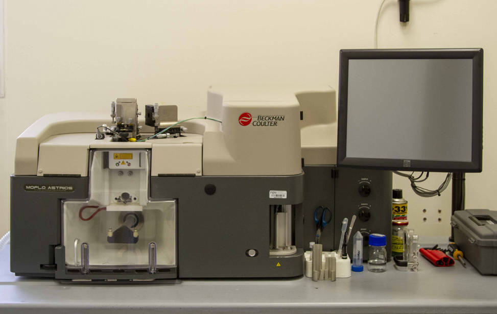
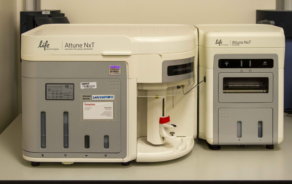
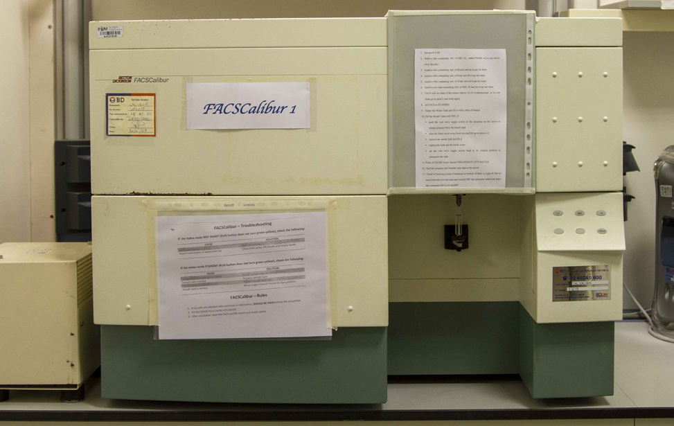
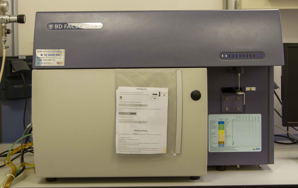
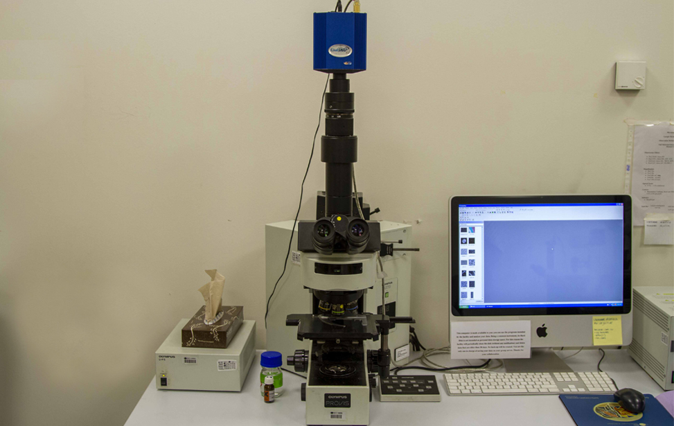
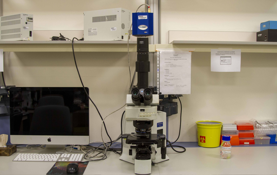
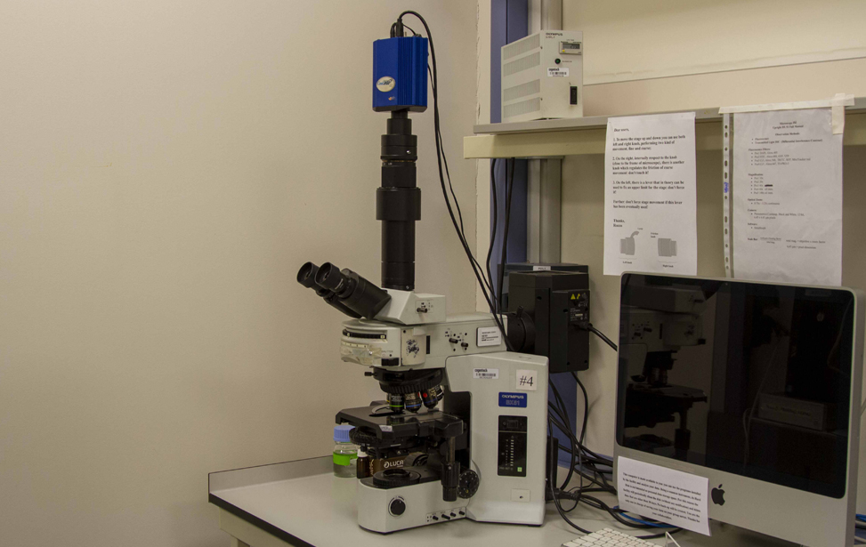
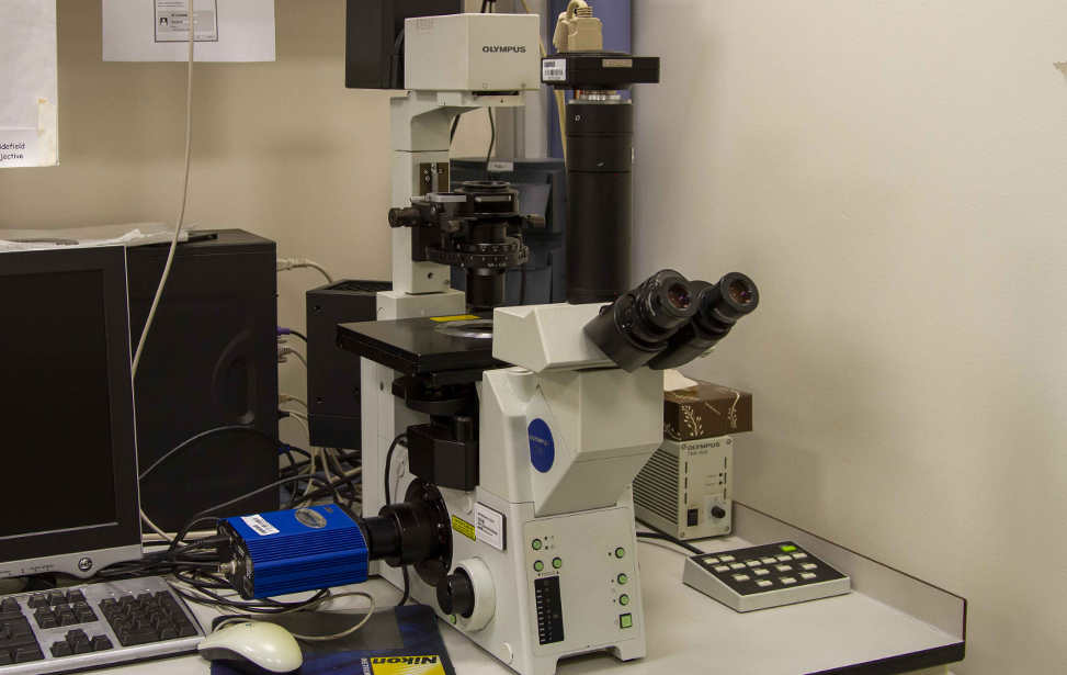
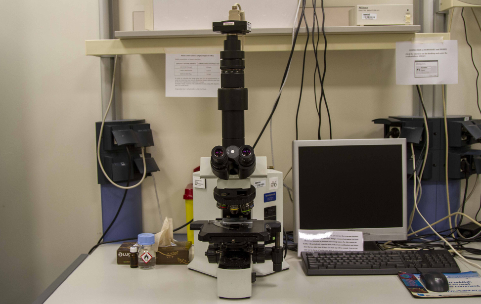
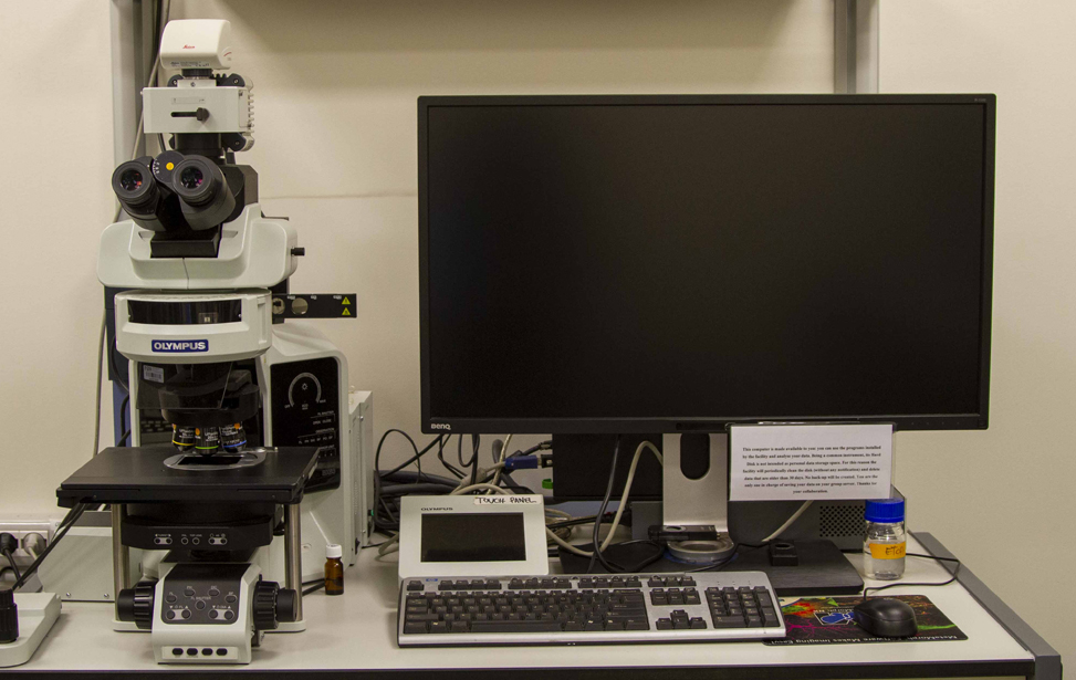
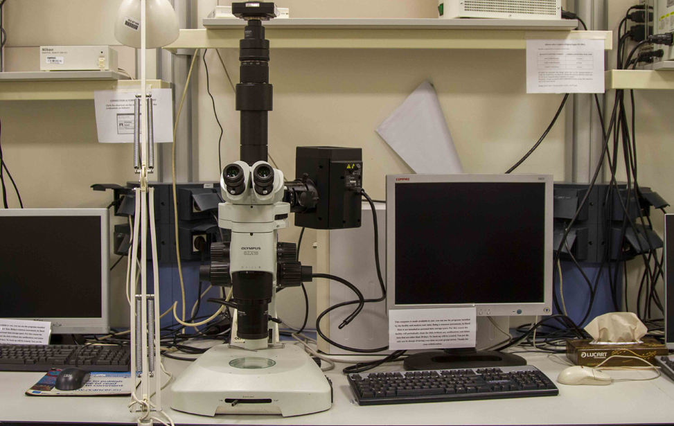
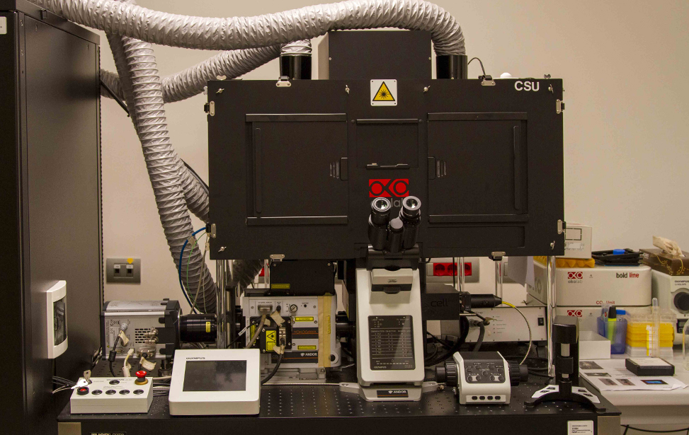
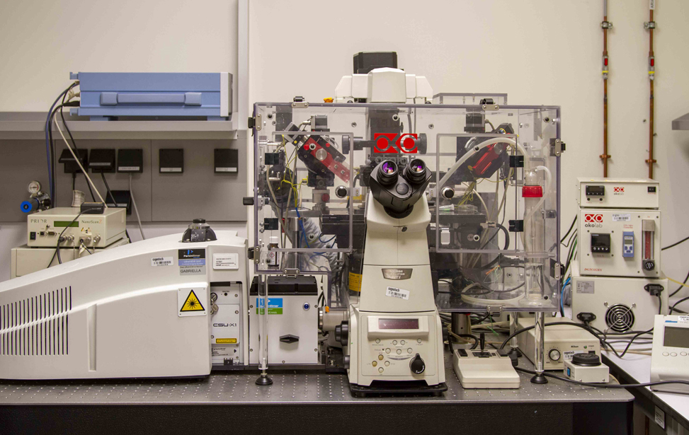
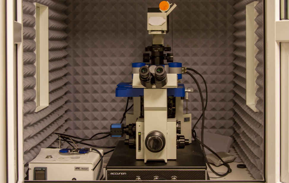
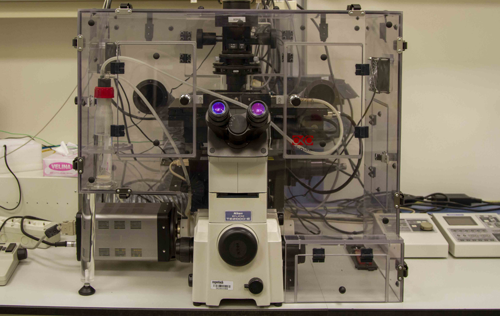
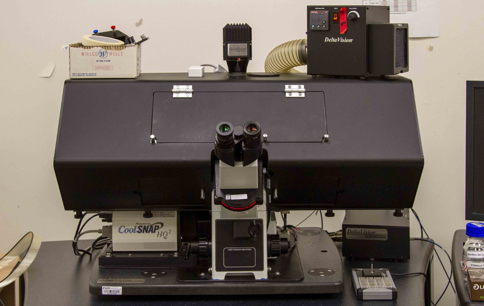
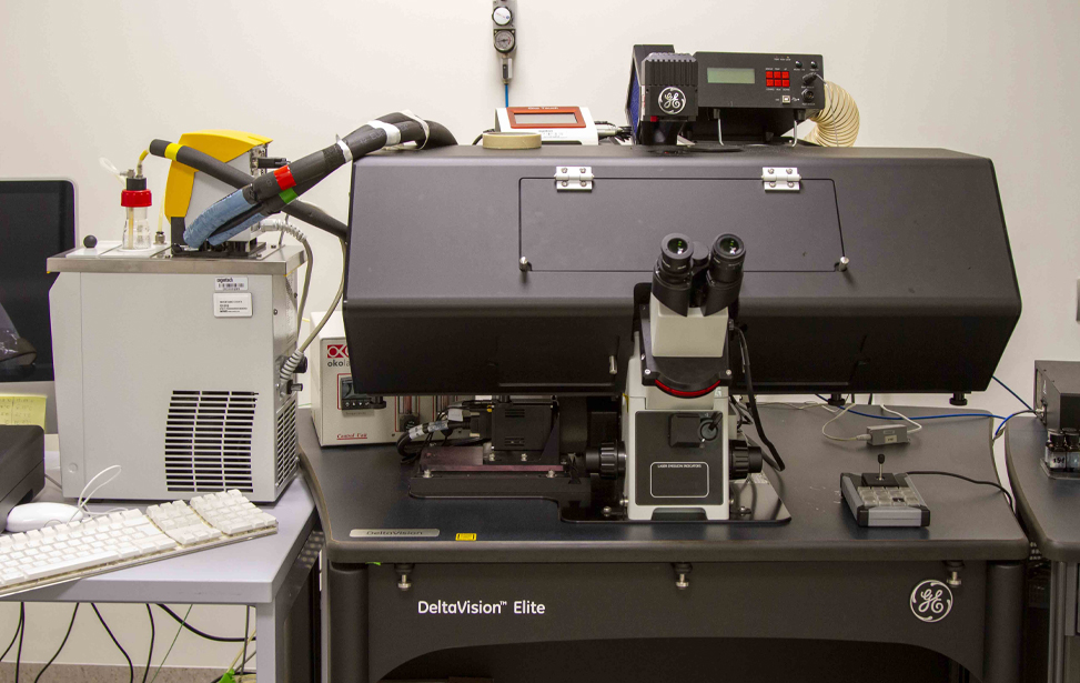
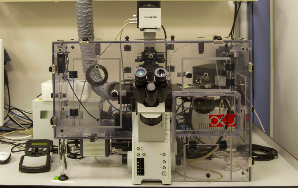
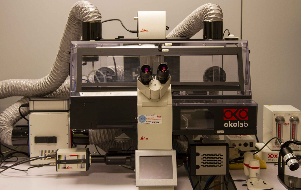
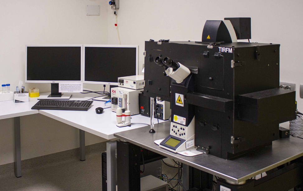
At IFOM, the Imaging Technological Development Unit (TDU) provides state-of-the-art technological platforms in the field of light microscopy, flow cytometry and cell sorting. A team of highly qualified and experienced people with different scientific backgrounds support IFOM's research activities, not only during the preparation and acquisition of the sample, but also in post-processing of data and image analysis.
The available systems meet the needs and demands of both senior researchers using sophisticated methods and technologies and students at the beginning of their scientific career who require assistance and dedicated training for professional development.
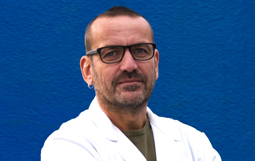
Dario Parazzoli
Born in Milano, Dario Parazzoli has shown very early his interest in the interplay of physics and biology. Graduated in Physics from the University in Milan with a thesis on the characterization of the dispersion pattern of radionuclides in the environment from the Chernobyl nuclear disaster, Dario's career has since then followed a path at the intersection of technology and the biological effects of radiations. Pursuing this passion, in 1997 he joined a leading industrial company in the field of microscopy where he had the opportunity to work as project manager at the forefront of confocal technology and high-end solutions for life science microscopy. In 2004, after almost a decade in the industry where he also refined his managerial abilities, Dario decided to deepen his interests in biomedical research by joining the research arm of the European Institute of Oncology in Milano where he was a key figure in setting up an advanced configuration of a Total Internal Fluorescence Microscope making it suitable for long time living cell applications. In 2008 he briefly joined the National Institute of Molecular Genetics where he established the imaging facility but in 2009 he was back in cancer research at IFOM where he now leads the Imaging Technological Development Unit (TDU).
Unit members
Sara BarozziShadi Bahiri
Francesca Casagrande
Zeno Lavagnino
Serena Magni
Sara Michela Martone
Fabrizio Orsenigo
Maria Grazia Totaro
Selected Unit Publications
- Kidiyoor GR, Li Q, Bastianello G, Bruhn C, Giovannetti I, Mohamood A, Beznoussenko GV, Mironov A, Raab M, Piel M, Restuccia U, Matafora V, Bachi A, Barozzi S, Parazzoli D, Frittoli E, Palamidessi A, Panciera T, Piccolo S, Scita G, Maiuri P, Havas KM, Zhou ZW, Kumar A, Bartek J, Wang ZQ, Foiani M. "ATR is essential for preservation of cell mechanics and nuclear integrity during interstitial migration.". Nat Commun. 2020 Sep 24;11(1):4828. doi: 10.1038/s41467-020-18580-9.PMID: 32973141 Free PMC article.
- Pessina F, Giavazzi F, Yin Y, Gioia U, Vitelli V, Galbiati A, Barozzi S, Garre M, Oldani A, Flaus A, Cerbino R, Parazzoli D, Rothenberg E, d'Adda di Fagagna F. "Functional transcription promoters at DNA double-strand breaks mediate RNA-driven phase separation of damage-response factors.". Nat Cell Biol. Nat Cell Biol. 2019 Oct;21(10):1286-1299. doi: 10.1038/s41556-019-0392-4. Epub 2019 Sep 30.PMID: 31570834 Free PMC article.
- Palamidessi A, Malinverno C, Frittoli E, Corallino S, Barbieri E, Sigismund S, Beznoussenko GV, Martini E, Garre M, Ferrara I, Tripodo C, Ascione F, Cavalcanti-Adam EA, Li Q, Di Fiore PP, Parazzoli D, Giavazzi F, Cerbino R, Scita G. "Unjamming overcomes kinetic and proliferation arrest in terminally differentiated cells and promotes collective motility of carcinoma.". Nat Mater. 2019 Nov;18(11):1252-1263. doi: 10.1038/s41563-019-0425-1. Epub 2019 Jul 22.PMID: 31332337 Free article.
- Malinverno C, Corallino S, Giavazzi F, Bergert M, Li Q, Leoni M, Disanza A, Frittoli E, Oldani A, Martini E, Lendenmann T, Deflorian G, Beznoussenko GV, Poulikakos D, Haur OK, Uroz M, Trepat X, Parazzoli D, Maiuri P, Yu W, Ferrari A, Cerbino R, Scita G. "Endocytic reawakening of motility in jammed epithelia." Nat Mater. 2017 May;16(5):587-596. doi: 10.1038/nmat4848. Epub 2017 Jan 30.PMID: 28135264 Free PMC article.
- Kumar A, Mazzanti M, Mistrik M, Kosar M, Beznoussenko GV, Mironov AA, Garrè M, Parazzoli D, Shivashankar GV, Scita G, Bartek J, Foiani M. "ATR mediates a checkpoint at the nuclear envelope in response to mechanical stress." Cell 2014 Jul 31;158(3):633-46. doi: 10.1016/j.cell.2014.05.046. PMID: 25083873 [pubmed - in process] Free PMC Article
- Frittoli E, Palamidessi A, Marighetti P, Confalonieri S, Bianchi F, Malinverno C, Mazzarol G, Viale G, Martin-Padura I, Garrè M, Parazzoli D, Mattei V, Cortellino S, Bertalot G, Di Fiore PP, Scita G. "A RAB5/RAB4 recycling circuitry induces a proteolytic invasive program and promotes tumor dissemination." J Cell Biol. 2014 Jul 21;206(2):307-28. doi: 10.1083/jcb.201403127. PMID: 25049275 [pubmed - indexed for MEDLINE]
- Palamidessi A, Frittoli E, Ducano N, Offenhauser N, Sigismund S, Kajiho H, Parazzoli D, Oldani A, Gobbi M, Serini G, Di Fiore PP, Scita G, Lanzetti L. "The GTPase-activating protein RN-tre controls focal adhesion turnover and cell migration." Curr Biol. 2013 Dec 2;23(23):2355-64. doi: 10.1016/j.cub.2013.09.060. Epub 2013 Nov 14. PMID: 24239119 [pubmed - indexed for MEDLINE] Free Article
- Meregalli M, Navarro C, Sitzia C, Farini A, Montani E, Wein N, Razini P, Beley C, Cassinelli L, Parolini D, Belicchi M, Parazzoli D, Garcia L, Torrente Y. "Full-length dysferlin expression driven by engineered human dystrophic blood derived CD133+ stem cells." FEBS J. 2013 Dec;280(23):6045-60. doi: 10.1111/febs.12523. Epub 2013 Oct 8. PMID: 24028392 [pubmed - indexed for MEDLINE]
- Singh AV, Vyas V, Montani E, Cartelli D, Parazzoli D, Oldani A, Zeri G, Orioli E, Gemmati D, Zamboni P. "Investigation of in vitro cytotoxicity of the redox state of ionic iron in neuroblastoma cells." J Neurosci Rural Pract. 2012 Sep;3(3):301-10. doi: 10.4103/0976-3147.102611. Erratum in: J Neurosci Rural Pract. 2013 Jan-Mar;4(1):98. Maontani, Erica [corrected to Montani, Erica]. PMID: 23188983 [pubmed] Free PMC Article
- Robert T, Vanoli F, Chiolo I, Shubassi G, Bernstein KA, Rothstein R, Botrugno OA, Parazzoli D, Oldani A, Minucci S, Foiani M. "HDACs link the DNA damage response, processing of double-strand breaks and autophagy." Nature 2011 Mar 3;471(7336):74-9. doi: 10.1038/nature09803. PMID: 21368826 [pubmed - indexed for MEDLINE] Free PMC Article
- Testa I, Parazzoli D, Barozzi S, Garrè M, Faretta M, Diaspro A. "Spatial control of pa-GFP photoactivation in living cells." J Microsc. 2008 Apr;230(Pt 1):48-60. doi: 10.1111/j.1365-2818.2008.01951.x. PMID: 18387039 [pubmed - indexed for MEDLINE]
- Testa I, Garrè M, Parazzoli D, Barozzi S, Ponzanelli I, Mazza D, Faretta M, Diaspro A. "Photoactivation of pa-GFP in 3D: optical tools for spatial confinement." Eur Biophys J. 2008 Sep;37(7):1219-27. doi: 10.1007/s00249-008-0317-9. Epub 2008 Apr 1. PMID: 18379772 [pubmed - indexed for MEDLINE]
- Diaspro A, Testa I, Faretta M, Magrassi R, Barozzi S, Parazzoli D, Vicidomini G. "3D localized photoactivation of pa-GFP in living cells using two-photon interactions." Conf Proc IEEE Eng Med Biol Soc. 2006;1:389-91. PMID: 17946398 [pubmed - indexed for MEDLINE]
- Corada M, Chimenti S, Cera MR, Vinci M, Salio M, Fiordaliso F, De Angelis N, Villa A, Bossi M, Staszewsky LI, Vecchi A, Parazzoli D, Motoike T, Latini R, Dejana E. "Junctional adhesion molecule-A-deficient polymorphonuclear cells show reduced diapedesis in peritonitis and heart ischemia-reperfusion injury." Proc Natl Acad Sci U S A. 2005 Jul 26;102(30):10634-9. Epub 2005 Jul 18. PMID: 16027360 [pubmed - indexed for MEDLINE] Free PMC Article
Photogallery
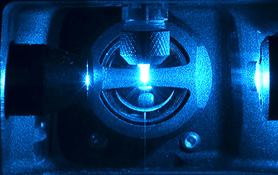
Figure 2: Interrogation point of a FACS Sorter

Figure 2: Particle Image Velocimetry (PIV) Analysis of a wound healing experiment
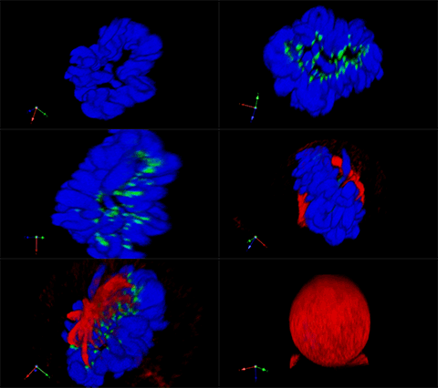
3D rendering of a mitotic He-La cell
(blue chromatin, green CREST, red tubulin)
